
Our Location
Monday
8:00 am - 5:00 pm
Tuesday
7:00 am - 5:00 pm
Wednesday
10:00 am - 7:00 pm
Thursday
8:00 am - 5:00 pm
Friday
8:00 am - 5:00 pm
Saturday
9:00 am - 2:00 pm*
*2 times per month
*2 times per month

Digital radiography replaces traditional film with electronic sensors that capture X-ray images and transmit them instantly to a computer. These sensors — often thin, flexible plates placed inside the mouth or larger detectors used for extraoral views — convert X-ray energy into digital data. That data is processed by specialized software, producing clear images that practitioners can view, enhance, and store with minimal delay. The result is a faster, more streamlined imaging process compared with film-based systems.
When an image is taken, it appears on the clinician’s monitor within seconds, which allows for immediate review and repeat imaging if needed. Modern software provides tools to adjust contrast, magnify areas of interest, and measure structures with precision. This immediate feedback helps reduce retakes and improves the efficiency of the appointment for both the patient and the clinical team.
Because images are captured digitally, they integrate seamlessly into electronic health records and imaging libraries. Digital files can be organized, archived, and retrieved without the physical storage demands of film. That organization supports long-term case management and makes it easier to compare images over time to monitor changes in oral health.
One of the most meaningful advantages of digital radiography is a reduction in radiation exposure compared with conventional film techniques. Advances in sensor sensitivity and imaging algorithms let clinicians obtain diagnostically useful images with lower X-ray doses. This makes digital radiography a safer choice for routine diagnostics while maintaining image quality that supports confident clinical decisions.
Digital sensors are also designed with patient comfort in mind. Many sensors are thinner and more ergonomic than older film packets, which can improve tolerance during intraoral imaging. Faster processing times mean patients spend less time holding sensors in place, and the likelihood of repeated exposures due to processing errors is greatly reduced.
From an environmental perspective, digital imaging eliminates the need for developing chemicals and film waste. By removing darkroom processes and chemical developers from the workflow, practices reduce hazardous waste and streamline their operations, which supports broader sustainability goals without compromising diagnostic capability.
High-resolution digital images enhance diagnostic accuracy by revealing fine anatomic detail that can be adjusted and analyzed in real time. Clinicians can zoom in on suspicious areas, fine-tune brightness and contrast, and apply measurement tools that assist in detecting decay, evaluating bone levels, and assessing root structure. These capabilities support earlier detection of problems and more precise treatment planning.
Digital radiography also facilitates interdisciplinary collaboration. Images can be shared securely with specialists, laboratories, or insurance providers as needed, accelerating consultations and treatment coordination. This connectivity is especially valuable for complex restorative and implant cases where clear visualization and shared planning are essential for predictable outcomes.
Because digital archives make comparing current and prior images straightforward, clinicians can monitor subtle changes over time. This historical perspective is invaluable for tracking disease progression, evaluating treatment response, and making informed decisions about ongoing preventive or restorative strategies.
Digital radiography is a foundational component of a digital dental workflow. It interoperates with intraoral cameras, digital impressions, and practice management systems to create a cohesive clinical environment. When images, treatment records, and digital impressions are available in the same system, the care team can coordinate more efficiently and present treatment options to patients with greater clarity.
For procedures such as implant planning or complex restorative work, digital imaging supports precise measurements and three-dimensional assessments when combined with advanced modalities. Even in routine care, the ability to display images on a monitor during an exam enhances communication: patients can see what the clinician sees, making it easier to explain findings and proposed treatments.
At Chadha & Co Dental, our use of digital radiography reflects a commitment to contemporary, evidence-informed dentistry. Integrating imaging into our practice workflow allows us to deliver care that is both efficient and patient-centered, while maintaining rigorous standards for recordkeeping and clinical documentation.
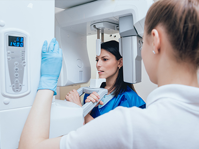
During a routine imaging appointment, a team member will position the sensor and take X-rays as directed by the clinician’s diagnostic needs. The process is typically quick and well-tolerated; intraoral images are captured in moments, and panoramic or extraoral views are completed in a single rotation. If you have concerns about sensor placement or comfort, the staff will take steps to minimize discomfort and ensure the images are obtained correctly the first time.
Safety remains a priority throughout the procedure. Lead aprons or thyroid collars may be used when appropriate, and exposure settings are adjusted to the lowest acceptable level for diagnostic quality. For children, expect tailored exposure techniques that reflect their smaller anatomy and increased sensitivity to radiation.
After images are captured, the clinician will review them with you and discuss any findings in plain language. You may see the images displayed on a monitor as the clinician explains what the images reveal and how they relate to oral health or treatment options. This shared review helps patients make informed decisions and understand the rationale behind recommended care.
In summary, digital radiography streamlines diagnosis, enhances safety, and supports clearer patient communication. If you’d like to learn more about how digital imaging is used in our office or how it might benefit your care, please contact us for more information.

Digital radiography is a dental imaging technique that uses electronic sensors to capture X-ray images instead of traditional film. The sensors convert X-ray energy into digital data that is processed by specialized software to produce clear, high-resolution images. These images can be viewed, enhanced, and stored instantly, which speeds up diagnosis and documentation compared with film-based systems.
Because the images are digital, they integrate with electronic health records and imaging libraries for easy retrieval and comparison. Clinicians can organize and archive files without the physical storage demands of film, supporting long-term case management. Digital files also reduce the need for chemical processing and physical handling, simplifying workflow and recordkeeping.
During digital radiography, an intraoral or extraoral sensor is positioned and a controlled X-ray exposure creates an electronic signal within the sensor. That signal is converted to a digital image by the sensor and immediately transmitted to a connected computer for processing. Software tools then allow clinicians to adjust contrast, magnify areas of interest, and perform measurements to aid in interpretation.
The immediate availability of images enables quick review and, when necessary, repeat imaging during the same appointment to ensure diagnostically useful views. Modern systems use algorithms to enhance image quality while minimizing noise, which supports accurate visualization of fine anatomic details. Integration with practice software means images can be associated directly with patient records for efficient documentation.
One key safety advantage of digital radiography is reduced radiation exposure compared with conventional film techniques, thanks to more sensitive sensors and improved image processing. Clinicians can obtain diagnostic-quality images at lower X-ray doses, which is beneficial for routine care and for patients who require frequent monitoring. Exposure settings are adjusted to the lowest level consistent with diagnostic needs, following the ALARA principle.
Digital systems also reduce the risk associated with processing errors that previously led to retakes, and modern sensor designs improve patient comfort to help minimize movement during exposures. Additional precautions such as thyroid collars and lead aprons are used when appropriate to further limit exposure to nearby tissues. For pediatric patients, clinicians apply age- and size-appropriate exposure protocols to account for greater sensitivity to radiation.
High-resolution digital images reveal fine anatomic details that support earlier and more accurate detection of conditions such as interproximal decay, bone loss, and root abnormalities. Clinicians can zoom in, adjust brightness and contrast, and use measurement tools to evaluate structures with precision, which enhances diagnostic confidence. The ability to compare current and prior images side by side also helps track progression or healing over time.
Digital images facilitate interdisciplinary collaboration by allowing secure electronic sharing with specialists, laboratories, and other members of the care team. For restorative and implant planning, these images can be incorporated into a broader digital workflow that includes digital impressions and three-dimensional imaging when required. The combined datasets support more predictable treatment sequences and clearer communication about clinical options.
During the appointment a team member will position a thin intraoral sensor or arrange an extraoral detector and take X-rays as directed by the clinician. The exposure itself is brief, and images typically appear on the clinician’s monitor within seconds for immediate review. Staff will take steps to maximize comfort, such as using small, ergonomic sensors and offering instructions to reduce movement during capture.
Safety measures, including lead aprons or thyroid collars, are used when appropriate and exposure settings are tailored to the diagnostic need. After images are captured the clinician will review them with you and explain any findings in plain language, showing the images on a screen when helpful. This shared review helps you understand the clinical rationale for recommended care and the next steps in treatment planning.
Digital radiography can be performed safely for children when clinicians follow pediatric exposure protocols that reduce dose and use appropriately sized sensors. Children receive customized settings to limit radiation while still producing diagnostically useful images, and staff use positioning aids and comfort measures to obtain clear results on the first attempt. Regular monitoring and preventive care planning often rely on these lower-dose techniques to minimize cumulative exposure.
For pregnant patients, dental radiographs are taken only when necessary and after discussion of the risks and benefits with the clinician. If imaging is required, extra precautions such as lead aprons and thyroid collars are routinely used to shield the abdomen and neck. Clinicians generally defer nonurgent radiographs until after pregnancy when possible and will document the rationale for any imaging performed during pregnancy.
Digital images are stored electronically within the practice’s imaging library and integrated patient records, which allows secure archiving and organized retrieval. Practices employ backup and access controls to protect patient data, and images are managed according to applicable privacy regulations and professional recordkeeping standards. Storing images digitally also eliminates chemical processing waste and physical film storage requirements.
When images need to be shared with specialists or laboratories, secure electronic transfer protocols are used to protect patient confidentiality. Clinicians typically include imaging in the clinical record so that previous and current images can be compared efficiently for ongoing care. Patients may request copies of their images as part of their records in accordance with standard medical record procedures.
Digital radiography is a foundational element of a cohesive digital dental workflow, interoperating with intraoral cameras, digital impression systems, and practice management software. When imaging, impressions, and treatment records are available in the same system, the care team can plan treatments more efficiently and coordinate steps such as restorations or implant placement. This interoperability supports clearer patient education by allowing clinicians to display combined visual information during consultations.
For complex procedures, digital radiographs can be paired with three-dimensional imaging and computer-aided planning tools to improve measurement accuracy and predictability. The combined digital datasets enable more precise surgical guides and restorative designs when indicated. Overall, the integrated approach reduces manual transfers and shortens the time between diagnosis and treatment execution.
Digital radiography is effective at identifying a range of dental conditions including interproximal cavities, periapical pathology, periodontal bone loss, and issues with root anatomy. High-resolution images allow clinicians to detect subtle changes in tooth structure and surrounding bone that might not be apparent on a visual exam alone. Serial images taken over time make it possible to observe trends such as lesion progression or bone level changes.
While two-dimensional digital radiographs are well suited for many diagnostic tasks, certain clinical situations may require additional modalities such as cone beam computed tomography for three-dimensional assessment. Clinicians will select the appropriate imaging technique based on the diagnostic question and the need for cross-sectional or volumetric detail. The chosen approach is documented and explained so patients understand why a specific modality is recommended.
At Chadha & Co Dental we use digital radiography to enhance diagnostic accuracy, improve patient safety, and streamline clinical workflow. The technology allows faster image acquisition, lower radiation doses, and immediate integration with the patient record, which supports efficient, evidence-informed care. Digital images also make it easier to communicate findings and treatment options with patients and with collaborating specialists.
Using digital imaging reduces the need for film processing and hazardous chemical waste, aligning imaging practices with modern sustainability and recordkeeping standards. The ability to store, compare, and securely share images contributes to consistent long-term monitoring and coordinated treatment planning. Overall, the technology supports a high standard of clinical documentation and patient-centered communication.
Ready to schedule your next dental appointment or have questions about our services?
Contacting Chadha & Co Dental is easy! Our friendly staff is available to assist you with scheduling appointments, answering inquiries about treatment options, and addressing any concerns you may have. Whether you prefer to give us a call, send us an email, or fill out our convenient online contact form, we're here to help. Don't wait to take the first step towards achieving the smile of your dreams – reach out to us today and discover the difference personalized dental care can make.
Back to top