
Our Location
Monday
8:00 am - 5:00 pm
Tuesday
7:00 am - 5:00 pm
Wednesday
10:00 am - 7:00 pm
Thursday
8:00 am - 5:00 pm
Friday
8:00 am - 5:00 pm
Saturday
9:00 am - 2:00 pm*
*2 times per month
*2 times per month

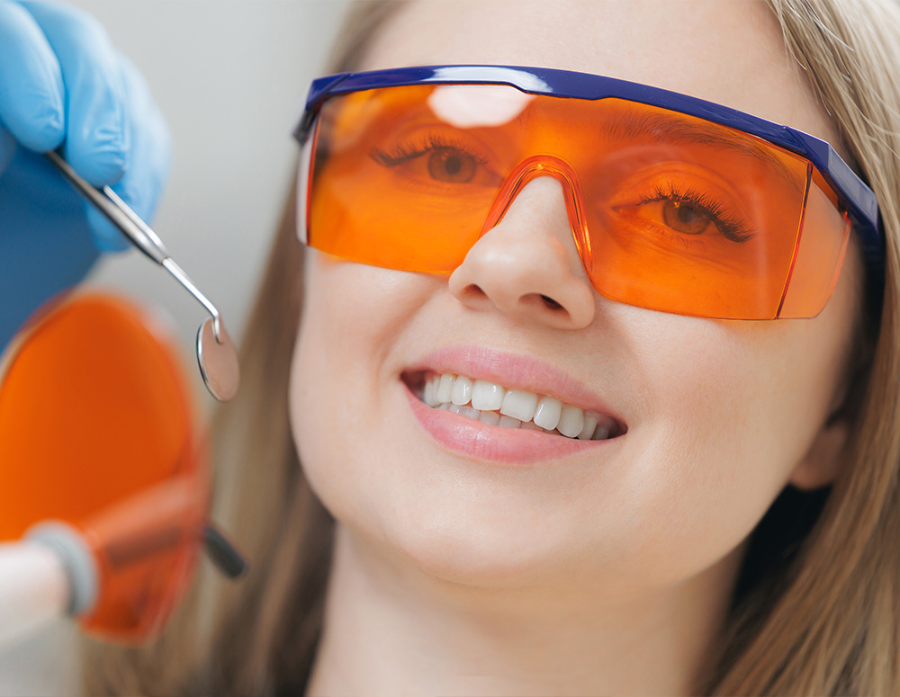
An intraoral camera is a small, pen-sized imaging device designed to capture detailed, full-color photographs and video inside the mouth. Unlike traditional dental mirrors, this tool provides close-up, high-resolution views of teeth, gums, and other oral structures. The live images are shown on a monitor so patients and clinicians can see the same perspective at the same time, making routine exams and more complex evaluations clearer and more precise.
Because the images are digital, they can be enlarged without loss of clarity, highlighted for emphasis, and saved into a patient’s chart. This ability to document the oral environment visually enhances diagnostic accuracy and creates a reliable record for monitoring changes over time. For patients, seeing an actual image often removes ambiguity and helps build understanding of a recommended course of care.
Clinically, intraoral cameras complement other diagnostic tools such as digital radiography and tactile examination. While X-rays reveal underlying structures like tooth roots and bone, intraoral images show surface anatomy, discoloration, micro-cracks, and early signs of wear or soft-tissue issues. Together, these resources create a fuller clinical picture that supports timely, evidence-based decision making.
At Chadha & Co Dental, we integrate intraoral imaging into routine assessments to make evaluations more transparent and collaborative. By combining visual documentation with professional interpretation, our team aims to catch problems earlier and to present findings to patients in a way that’s easy to understand.
One of the most important benefits of intraoral imaging is its impact on diagnostic clarity. Small defects, hairline cracks, early decay between teeth, and areas of abnormal soft tissue can be challenging to detect with the naked eye alone. The magnified, well-lit view produced by an intraoral camera often reveals subtle changes that might otherwise be missed, allowing clinicians to prioritize care based on objective visual evidence.
Saved images become part of the patient record and can be compared over time to monitor progression or healing. This longitudinal perspective is especially valuable for managing chronic conditions such as periodontal disease, for tracking the effectiveness of restorations, and for documenting pre- and post-treatment status. Clear visual records help the dental team refine treatment plans and sequence care efficiently.
In treatment planning, intraoral photographs aid in communication among the dentist, specialists, and the dental laboratory. When a restoration, implant, or periodontal procedure is under consideration, precise images help convey shape, color, and anatomical relationships. This reduces ambiguity when fabricating crowns, bridges, or other prosthetics, and supports more predictable results.
Visual information changes the way patients participate in their care. When clinicians can show real-time images of a questionable area, patients gain immediate insight into what is happening in their own mouths. This shared view fosters a collaborative dialogue, enabling patients to ask targeted questions and to weigh options with a clearer sense of the potential benefits and risks.
Informed consent is strengthened when recommendations are supported by images a patient can see and keep in their chart. Rather than relying solely on verbal descriptions or clinical jargon, patients can review photographs that illustrate why a particular intervention is advised. This transparency often increases confidence in the proposed plan and reduces confusion about next steps.
For families and caregivers, intraoral images provide a straightforward way to explain treatment needs, especially for children or patients who may be anxious. The visual approach is accessible and respectful of patient intelligence, helping people understand both immediate concerns and long-term maintenance priorities without oversimplification.
Because communication is at the heart of quality dental care, our team uses intraoral photos as a routine educational tool. It helps ensure that patients are fully informed and actively involved in decisions about their oral health.
Modern intraoral cameras produce files that integrate seamlessly with practice management and imaging software. Photographs can be attached to patient charts, annotated, and shared securely with other clinicians when specialty care or multi-disciplinary consultation is needed. This digital compatibility streamlines referrals and reduces delays in coordinating complex care.
When collaborating with specialists — for example, periodontists, endodontists, or prosthodontists — high-quality intraoral images provide a common visual language. Specialists can review the exact conditions seen in the patient’s mouth before they ever arrive in the consult room, allowing for more focused recommendations and efficient use of appointment time.
Dental laboratories also benefit from precise intraoral photos. When fabricating custom restorations, lab technicians use images to match shade, morphology, and occlusal relationships more accurately, which often reduces the need for multiple adjustments. That level of detail contributes to restorations that fit better and look more natural, improving the overall patient experience.
Intraoral cameras are designed for frequent clinical use and are incorporated into infection control protocols. Most devices use disposable sheaths or autoclavable tips to maintain sterility between patients, while the handheld components are cleaned and disinfected according to manufacturer guidelines and industry standards. These practices ensure imaging contributes to care without compromising safety.
Clinicians receive training in proper camera handling, lighting control, and image capture techniques to maximize clarity and minimize discomfort. Good technique includes steady positioning, appropriate angulation, and consistent lighting, all of which improve the diagnostic value of each image. Training also covers efficient file management so images are stored accurately in the electronic chart.
Regular maintenance and software updates keep intraoral imaging systems functioning reliably. Calibration, careful storage, and adherence to recommended cleaning procedures extend device longevity and preserve image quality. In the clinical setting, these operational details matter: reliable technology supports consistent, high-quality patient care.
By following these best practices, dental teams can incorporate intraoral imaging as a routine, dependable part of examinations and procedures, enhancing both safety and diagnostic yield.
Conclusion: Intraoral imaging makes dental care more transparent, accurate, and patient-centered. By capturing high-resolution photos and video, clinicians can identify subtle problems earlier, build clearer treatment plans, and involve patients more directly in decisions about their care. At Chadha & Co Dental we use intraoral cameras as part of a coordinated, modern approach to oral health that emphasizes clarity and collaboration. If you’d like to learn more about how intraoral imaging is used during exams and treatments, please contact us for additional information.

An intraoral camera is a small, pen-sized imaging device that captures high-resolution, full-color photographs and short video from inside the mouth. The device uses a tiny lens and LED illumination to produce magnified, well-lit views of teeth, gums, and other oral structures that are difficult to see with the naked eye. Live images are displayed on a monitor so clinicians and patients can view the same perspective in real time.
Because the images are digital they can be enlarged, annotated, and saved to the electronic health record for ongoing comparison. This visual documentation supports more accurate diagnosis and allows clinicians to monitor changes over time. The practice integrates intraoral imaging into routine exams to improve transparency and patient understanding.
Intraoral cameras reveal surface details such as hairline cracks, early interproximal decay, discoloration, and soft-tissue irregularities that are often difficult to detect with a mirror or visual inspection alone. Magnification, consistent lighting, and close proximity increase the chance of identifying subtle problems at an earlier stage. These images complement tactile examination and reduce diagnostic uncertainty.
Captured photographs become part of the patient record and permit direct comparison across visits, which is valuable for monitoring periodontal conditions, the integrity of restorations, and progression of lesions. This longitudinal perspective helps clinicians prioritize care based on objective visual evidence. By combining intraoral images with other diagnostic tools, providers can make more informed, evidence-based recommendations.
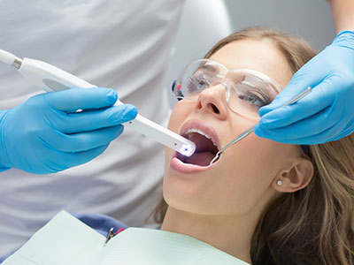
An intraoral camera exam is quick and noninvasive and is typically performed during a routine checkup or when a specific concern arises. The clinician or hygienist guides the handheld camera to capture still images and short video clips while the patient remains seated comfortably in the dental chair. Most patients experience no discomfort beyond minor tongue or lip movement as the device is positioned.
Images are displayed immediately on a monitor so the clinician can point out areas of interest and explain findings in plain language. Photographs may be annotated and saved to the chart for later review or comparison at follow-up visits. This process helps patients understand clinical recommendations and participate more fully in treatment decisions.
Intraoral cameras use visible light and pose no radiation risk, making them safe for repeated clinical use. Devices are used with disposable sheaths or autoclavable tips and are cleaned and disinfected according to manufacturer guidance to meet infection-control standards. Clinicians trained in proper handling minimize patient discomfort and ensure consistent image quality.
Digital files generated by intraoral cameras are stored in secure practice management systems and are protected under the same privacy practices that govern the rest of the patient record. When images are shared with specialists or laboratories they are transmitted using secure, HIPAA-compliant channels to maintain confidentiality. Patients may request copies of their images as part of their health record through standard office procedures.
High-quality intraoral photographs convey precise information about tooth shape, shade, and occlusal relationships that directly inform restorative planning. These images help the dental team and laboratory technicians visualize the exact conditions that restorations must match, reducing ambiguity during fabrication. Clear visual references improve the predictability and aesthetics of crowns, bridges, veneers, and implant restorations.
When planning complex care, clinicians combine intraoral images with radiographs and digital impressions to sequence treatment logically and set realistic expectations. Visual documentation also facilitates communication among the dentist, specialists, and the dental laboratory, which helps avoid unnecessary adjustments. Together, these resources contribute to more efficient appointments and higher-quality outcomes.
Yes. Intraoral camera files integrate with imaging and practice management software, making it straightforward to attach photographs to a patient’s chart for referral or consultation. Secure digital sharing enables specialists to review the mouth's visual condition before an in-person consultation, which can streamline referrals and focus the specialist's evaluation. This pre-review often leads to more targeted recommendations and efficient use of appointment time.
Dental laboratories use detailed photos to match shade, contour, and occlusal relationships when fabricating custom prosthetics, which can reduce the need for multiple remakes. When photos are accompanied by precise clinical notes and digital impressions, lab technicians can produce restorations that blend seamlessly with the patient’s natural dentition. Secure workflows preserve patient confidentiality while supporting close collaboration.
No. Intraoral cameras do not replace radiographs; they complement radiographic imaging by showing surface anatomy and soft-tissue detail that X-rays cannot display. Radiographs remain essential for evaluating root structure, bone levels, and interproximal decay beneath contact points. Together, images and X-rays provide a more complete diagnostic picture than either tool alone.
Clinicians use both modalities to cross-reference findings and to confirm diagnoses before recommending treatment. For example, a suspicious surface lesion identified with an intraoral camera may prompt targeted radiographs or additional testing to assess underlying structures. This combined approach improves diagnostic confidence and helps tailor treatment to the patient’s needs.
Visual evidence from intraoral cameras makes clinical explanations more accessible and helps patients grasp the nature and extent of oral health issues. Seeing the condition firsthand reduces ambiguity and enables more meaningful conversations about risks, benefits, and alternatives. This shared understanding supports informed decision-making and strengthens the informed-consent process.
Clinicians can annotate images to highlight areas of concern and to illustrate expected outcomes of proposed treatments, which helps set realistic expectations. Families and caregivers also benefit from clear visual records when coordinating care for children or patients with special needs. Routine use of intraoral imaging fosters transparency and encourages patients to take an active role in preventive care.
Proper infection-control measures are central to using intraoral cameras safely in a clinical setting, including single-use disposable sheaths or sterilizable tips for patient-to-patient protection. Handheld components are cleaned and disinfected following manufacturer instructions and industry guidelines to prevent cross-contamination. Staff training emphasizes consistent technique for image capture and strict adherence to sterilization protocols.
Regular maintenance, software updates, and periodic calibration preserve image quality and extend device lifespan. The practice schedules routine checks and follows recommended storage and handling procedures to avoid damage and downtime. Reliable equipment and disciplined maintenance protocols contribute to consistent diagnostic performance.
Chadha & Co Dental integrates intraoral imaging into routine exams to make evaluations more transparent, collaborative, and accurate. The technology supports early detection of issues, clear treatment planning, and more meaningful patient education, which aligns with the practice’s emphasis on comprehensive, patient-centered care. Using visual documentation alongside other digital tools helps the team deliver predictable, high-quality results.
Patients at our North Bethesda office can expect images to be included in their electronic chart and used to guide conversations during visits and follow-up care. When specialist referral or laboratory work is required, intraoral photographs help ensure that communication is precise and efficient. Those who want to learn more about how we use intraoral imaging during exams are encouraged to contact the office for a discussion.
Ready to schedule your next dental appointment or have questions about our services?
Contacting Chadha & Co Dental is easy! Our friendly staff is available to assist you with scheduling appointments, answering inquiries about treatment options, and addressing any concerns you may have. Whether you prefer to give us a call, send us an email, or fill out our convenient online contact form, we're here to help. Don't wait to take the first step towards achieving the smile of your dreams – reach out to us today and discover the difference personalized dental care can make.
Back to top